Nano-liter Microfluidic device

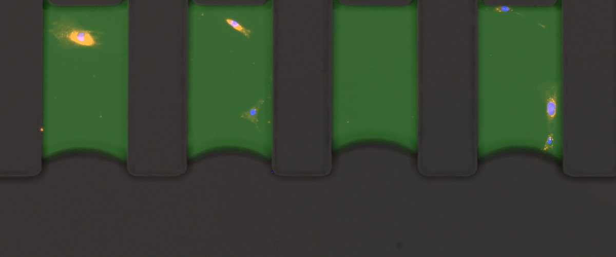
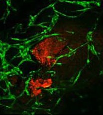
AST (Antimicrobial susceptibility testing ) system on a microfluidic device capable of delivering results in under 6 hours was developed in the Levenberg’s lab. Using an automated algorithm for data analysis, the system is capable of determining whether bacteria from an infection site is resistant or susceptible to the tested antibiotic. This technology was licensed to Nanosynex, and now-a-days, Nanosynex takes this proof of concept from the lab to the market.

Naosynex

Aleph Pharm Steak

Cell-Grown Meat

The aim of this project is to produce meat from cell cultures, using tissue engineering techniques. We isolate stem cells from a bovine origin, expand them, and seed them on 3D scaffolds, to develop bovine skeletal muscle tissues.
We study food-related properties of bovine skeletal muscle tissues under different cell combinations, scaffolds and media composition.

This study is supported by Aleph Frams (Aleph Farms)

Michigan fibers

Effect of scaffold geometry on vascular networks

Many environmental factors can affect the behavior of newly sprouting vessels. Among them, scaffold geometry provides mechanical cues that are translated into chemical and physical cell signaling, which impacts on the decisions made by migrating vessels. This research studies how different scaffold geometries can help control the behavior and orientation of sprouting vessels during network formation.

This research is supported by funding from the University of Michigan – Israel Partnership for Research.


Alignment of engineered blood vessels due to mechanical stimulation

Geometric and Mechanical Patterning of Hierarchical Vascular Networks

This research focuses on exploring the biophysical factors controlling vascular architecture, remodeling, and integration upon implantation. Using cutting edge methods to pattern the organization of cells, and to map and control mechanical forces within the 3D constructs, the role of cell-generated forces, initial endothelial cell organization and external mechanical forces in the location, orientation and extent of sprouting is studied. In addition, the impact of hierarchical network geometry, rationally designed, on implant integration in-vivo is assessed.

This research is supported by United States-Israel BSF grant in collaboration with Prof. Christopher Chen from Boston University.

cardiomyocytes

Cardiac Muscle Regeneration using a Perfusable Cardiac Patch

The aim of this project is to create a perfusable cardiac patch, intended for repairing damaged areas of the cardiac muscle. The patch is created using 3D bioprinting technology, which allows for the controlled and automatic deposition of biological and structural materials that will form the construct. Endothelial cells are deposited within an extracellular matrix mimicking material, enabling them to spontaneously organize into vascular networks. Cardiomyocytes are obtained by differentiating induced pluripotent stem cells (iPSCs) and seeded along the endothelial cells, creating a more complex tissue.

Spinal cord injured rat after exosome treatment

Spinal Cord Injury Regeneration

Stem cell-based therapies hold great potential to treat spinal cord injury and additional nervous system traumatic syndromes due to lack of regeneration in the adult nervous system. Our research focuses on delivering stem cells into a complete spinal cord injury. We utilize tissue engineering methods to enhance stem cell integration and survival post implantation. In addition we study the effect of vascularization on spinal cord regeneration. Alternatively, we exploit the regenerative effects of stem cell-derived exosomes as therapeutics and drug delivery platforms to target the spinal cord lesion.

This study was supported by the J&J Shervington Fund (SL), and the Israel Foundation for Spinal Cord Injury (SL).


Bone Tissue Repair

The main hurdle in bone tissue repair is maintaining appropriate vascularization for regeneration of large-scaled defects. We fabricate engineered constructs, capable of soft and hard tissue repair. These pre-vascularized composite grafts are fabricated from FDA approved and biocompatable biomaterials, which undergo an induction phase by differential seeding with diverse cellular components. Our bone tissue engineering (BTE) models are treated from a clinical perspective, utilizing in vivo imaging and CAD-CAM interfaces to produce defect-specific grafts. These engineered constructs are implanted in live animal models to assess their ability to regenerate complex anatomical structures.

This study is supported by the Israel science foundation.

muscle rita

Restoring Insulin Sensitivity in Type 2 Diabetes Patients using Engineered Muscle Tissue

Type 2 Diabetes (DM2) is a complex metabolic disease, characterized by adipose and muscle insulin resistance accompanied by defects in pancreatic insulin secretion or loss of function of insulin-secreting cells. Present therapeutic modalities include lifestyle modification and pharmaceutical agents, however many patients fail to achieve blood glucose homeostasis.
This research proposes to overcome peripheral tissue insulin resistance by genetically modifying skeletal muscle cells and use them to construct engineered muscle tissue. Upon implantation of such engineered muscle, overall glucose uptake of the animal is expected to be enhanced, therefore improving diabetic state.

This study is supported by the The Rina and Avner Schneur Center of Diabetes Research (http://schneur-diabetic-center.net.technion.ac.il/)

3D Bioprinting of Pancreatic Tissue

Developing therapies for pancreatic diseases, such as diabetes and pancreatic cancer, is hampered by a limited access to pancreatic tissue in vivo. Engineering three-dimensional (3D) tissue models, which accurately mimic the native organ, have great potential in biomedical applications, by both providing powerful platforms for studying tissue development and homeostasis and for modeling diseases in pharmaceutical testing.

Our research establishes a multi-disciplinary European consortium with the goal of developing an innovative bio-printing approach for generating pancreatic tissue. Tissues and organs comprise multiple cell types with specific biological functions that must be recapitulated in the printed tissue. We aim at bio-mimicking developmental processes to fabricate 3D bio-printed pancreatic tissue units that allow sustained cell viability, expansion and functional differentiation ex vivo.

Engineering  blood vessel networks

In this project we explored the effects of mechanical forces, scaffold type and supporting cells on angiogenesis. We examined different endothelial cells and support cells under various conditions, with a particular focus on  how engineered vessels align in response to mechanical forces, and  integrate in-vivo.